Disha nagari, behind master cook hotel, beed by pass road, chh. Sambhaji nagar.

shripadjoshi2007@gmail.com

+91 9307244003 /7058043743

ACL Reconstruction

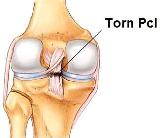
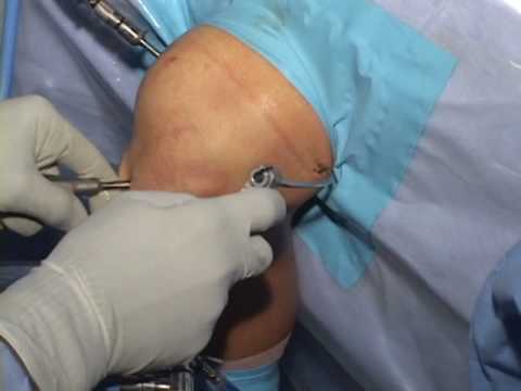
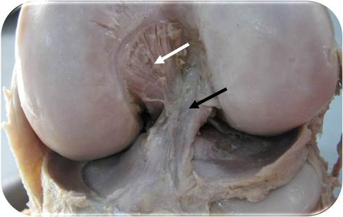
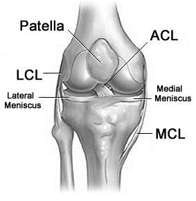
ACL reconstruction is surgery to reconstruct the ligament in the center of your knee. The anterior cruciate ligament (ACL) keeps your shin bone (tibia) in place. A tear of this ligament can cause your knee to give way during physical activity.Anterior cruciate ligament reconstruction (ACL reconstruction) is a surgical tissue graft replacement of the anterior cruciate ligament located in the knee, to restore its function after an injury. The torn ligament is removed from the knee before the graft is inserted in an arthroscopic procedure.The knee is essentially a hinged joint that is held together by the medial collateral (MCL), lateral collateral (LCL), anterior cruciate (ACL) and posterior cruciate (PCL) ligaments. The ACL runs diagonally in the middle of the knee, preventing the tibia from sliding out in front of the femur as well as providing rotational stability to the knee.


PCL Reconstruction

The function of the PCL is to prevent the femur from sliding off the anterior edge of the tibia and to prevent the tibia from displacing posterior to the femur. The posterior cruciate ligament is located within the knee.Severe PCL laxity, which results in a knee with significant posterior translation is quite unsettling to the patient, especially athletes due to the shifting of the tibia during running. These patients benefit from a PCL reconstruction which re-establishes stability to the knee. As noted in the treatment of ACL injuries the ruptured ligament cannot be repaired.  It must be reconstructed using a graft.


PLC Reconstruction

The PLC is comprised of multiple ligamentous and tendinous structures around the knee. A tendon represents connect tissue that attaches the muscle to the bone while a ligament is fibrous tissue that connects one bone to another. The PLC is located on the outside back (posterolateral) corner of the knee. Though many structures comprise the PLC, the main structures include the biceps femoris muscle and tendon (outside hamstring muscle), lateral collateral ligament popliteus muscle and tendon, popliteofibular ligament, fabellofibular ligament, arcuate ligament complex, and posterolateral joint capsule.

MCL Reconstruction

The medial collateral ligament (MCL) is a wide thick band of tissue running down the inner knee from the thighbone (femur) to a point on the shinbone (tibia) about four to six inches from the knee.Medial collateral ligament (MCL) reconstruction have been associated with extensive exposure risk of donor site morbidity with autografts, loss of motion, nonanatomic graft placement, and technical complexity with double-bundle constructs. Therefore, we implemented a technique that uses Achilles allograft, small incisions, and anatomic insertions to reconstruct the MCL.

The MCL’s main function is to prevent the leg from extending too far inward, but it also helps keep the knee stable and allows it to rotate.

We are here to help you Get relief from your pain

Get reed of from various body pain from our experienced doctor.

Scroll to Top